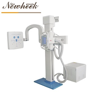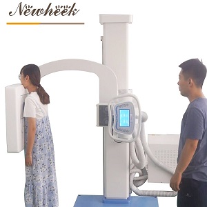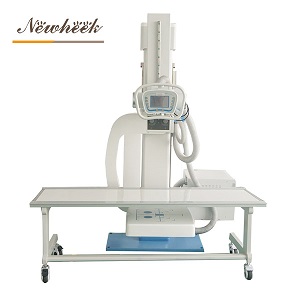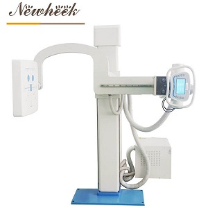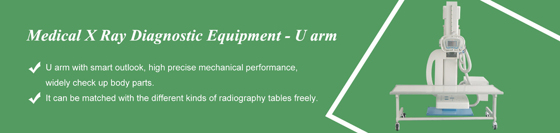

Quick answer: a U-arm DR is a digital radiography system in which the X-ray tube and the flat-panel detector are carried on the opposite ends of one U-shaped arm, powered by a high-frequency generator, and installed as fixed equipment in a dedicated radiography room. Every word in that long product name is doing real work — U-arm describes how the machine moves, digital describes how the image is captured, high-frequency describes how the X-ray output is produced, and fixed describes how it lives in your room. Buyers who understand each word separately negotiate better, install faster, and end up with a system that actually matches their caseload. This article decodes the name word by word.

Strip away the electronics and a U-arm DR is, at heart, one mechanical idea: the X-ray tube and the image receptor travel together on the same rigid, U-shaped arm. The tube sits at one end, the flat-panel detector at the other, and the arm pivots around a central column. That single design fact drives most of the day-to-day advantages.
On older room layouts, the tube head and the bucky or table had to be lined up by hand for every exposure — a small misalignment meant a clipped image or a repeat. On a U-arm, tube and detector are born aligned and stay aligned, because they are physically the same structure. When the operator raises the arm, both sides rise together. When it rotates, both sides rotate together. There is simply no alignment step to get wrong, which is one of the quietest but most valuable features of the design in a busy room.
Because tube and detector move as one unit, the operator can position the patient once — standing, seated, or lying — and then bring the arm to the patient. A chest exam on a standing patient, a frontal view on someone in a wheelchair, a reclined study on the table: in each case the arm’s vertical travel and rotation cover the change of angle, not the patient. For anyone who has ever tried to reposition an unwell or elderly patient four times for four views, this is the difference that sells the machine.
The geometry has honest limits, of course. The arm’s rotation path is built around frontal and near-frontal work; for true lateral, oblique, or cross-table geometries you need to understand exactly what the arm can and cannot reach — we break down those angled-view limits separately, because they matter more in orthopedic and trauma practice than most listings admit.
A U-arm DR concentrates the tube, the detector, the collimator, and their drives into a single floor-mounted column. Compare that with a classic room built from a tube stand, a wall bucky, and a table as three separate track-mounted pieces. For clinics converting a consulting room or a small annex into an imaging room, the single-column footprint is usually what makes the project feasible at all. The full list of what the room itself must provide — space, ceiling height, floor, power, shielding — is covered in our room requirements guide, and it is worth reading before you fall in love with any machine.
The DR in the name stands for digital radiography, and the practical meaning is simple: the image is captured by a flat-panel detector and appears on a workstation within seconds, with no film and no processing step in between.
No consumables. Film and chemicals are gone. There is nothing to reorder, nothing to store, nothing to expire in a cabinet. For facilities in regions where film supply is unreliable or expensive, this alone changes the economics of running the room.
No waiting. The preview appears right after the exposure. If the positioning was wrong, the patient is still on the spot and the repeat costs seconds, not a second appointment. Throughput and patient experience both improve for the same reason.
Images that travel. A digital image can be sent to a reviewing workstation, archived, attached to a report, or shared with an off-site radiologist. Film had to be physically carried; a digital file does not.
It is worth distinguishing DR from computed radiography (CR), the halfway technology that scans a phosphor plate after exposure. CR is digital too, but it keeps a physical cassette in the loop and adds a plate-handling step between exposure and image. A flat-panel DR system removes that step entirely. If you are weighing the two paths, our comparison of analog and digital X-ray imaging walks through the older technologies in more detail, and this overview of traditional versus digital imaging covers what changes for the staff, not just the hardware.
One honest note on scope: the detector is a component with its own deep vocabulary — panel sizes, scintillator types, pixel characteristics. Those internals deserve their own reading and are covered in depth on our detector-focused sister resource; in the buying conversation, what matters most at the machine level is confirming which detector is paired with the arm and that it integrates cleanly with the workstation software.
The generator is the part of the machine that takes wall power and turns it into the precisely controlled electrical supply the X-ray tube needs. High-frequency describes the modern way of doing that: instead of drawing power in large, slow pulses the way older generator designs did, a high-frequency generator switches its supply thousands of times per second and regulates the tube output continuously.
Why should a buyer care? Three practical reasons:
Reproducible exposures. When the generator holds the output steady, the 200th chest film of the week looks like the first. Consistency is what makes follow-up comparisons meaningful and what keeps repeat rates low.
A compact console. High-frequency designs need far less iron and copper to do the same job, so the generator cabinet and the control console shrink. In a small room — which, as discussed, is exactly the kind of room a U-arm is chosen for — that compactness is not cosmetic.
Quieter, cleaner power behavior on the room’s electrical supply, which matters in clinics where the imaging room shares its feed with other equipment.
When you compare offers, ask what generator class each machine carries and what the console actually controls — technique selection, anatomical presets, exposure timing. Two machines can look identical in a photo and differ completely in how the operator experiences a working day.
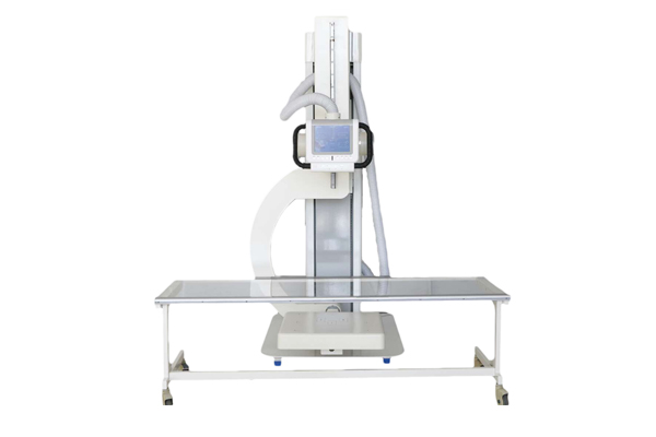
The final word in the name tells you how the machine lives. A fixed U-arm DR is floor-mounted, anchored, calibrated, and stays in its room for its whole working life. That is the opposite end of the spectrum from a mobile DR unit, which trades stability for the ability to travel to the patient’s bedside.
Stability you can build protocols on. Because nothing on a fixed system is designed to be pushed through corridors, the machine can be heavier, more rigid, and more precisely calibrated. Exposures are repeatable; the geometry does not drift; the room can be shielded once, correctly, around a machine that will not move.
Throughput. A fixed room is a production line: patients come to it, one after another, and the workflow — register, position, expose, review — compounds over a day. Mobile units shine at the bedside, but nobody processes a high daily caseload on one.
A dedicated, controlled environment. The room’s lighting, power, and radiation shielding are designed around the machine. That is better for image quality, better for staff, and better for compliance.
You need a room, and the room needs to qualify. Power capacity, floor condition, ceiling height, shielding — the machine is only as installable as the room it lands in, which is why we published a pre-shipment room checklist alongside our detailed requirements guide. If your use case genuinely requires imaging at the patient’s location — home visits, wards without an imaging room, multi-site clinics — then a mobile or portable device is the right category, and pretending a fixed system will cover it serves nobody. The two categories are complements, not competitors, and the cost relationship between mobile and fixed DR is worth understanding before budgeting either one.
Put the words back together and the machine’s working range becomes clear:
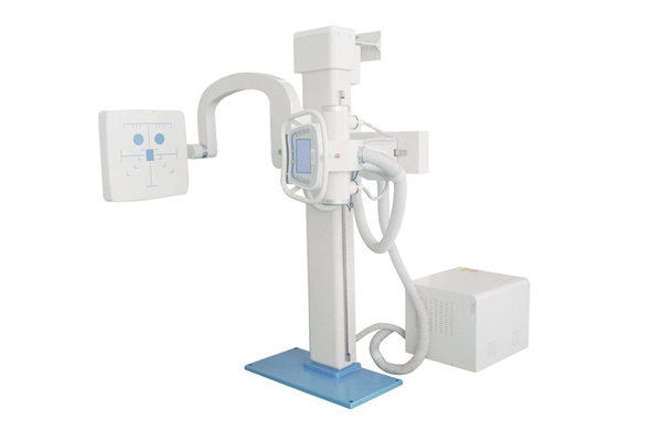
A responsible overview ends with the boundaries, because the most expensive mistake in imaging purchases is buying the wrong category confidently.
It is not a fluoroscopy or angiography machine. A U-arm DR captures still images — excellent ones — but it does not provide the continuous real-time imaging that guided procedures require. If your caseload includes contrast studies, interventional guidance, or any work where you need to watch motion on a monitor in real time, that is C-arm territory, and the two machines coexist in hospitals for exactly that reason. Our U-arm versus C-arm comparison draws that line carefully.
It is not a high-throughput, double-column main-frame. Large hospitals running two rooms’ worth of chest and skeletal volume often standardize on double-column systems with higher duty cycles. The U-arm DR wins on footprint, flexibility, and cost efficiency — the scenarios where it genuinely earns its keep are mapped room by room here — but a purchase decision should start from caseload, not from machine aesthetics.
It is not angle-free, but its angles have limits. As noted above, frontal and near-frontal projections are home turf; steep oblique and cross-table geometries need a frank conversation before purchase.
Armed with the decoded name, a buyer can interrogate any quotation — ours or a competitor’s — in four questions:
When the machine does arrive, the first 48 hours of unpacking and inspection set the tone for the whole installation — here is what to check when the crate arrives, so that any transit damage is documented while the claim window is open.
The names are used interchangeably across the industry, and most listings — including ours — mean the same machine: a single U-shaped arm carrying tube and detector on one shared structure. There are regional and vendor habits in the naming, and our U-arm versus UC-arm explainer untangles the terminology so you can compare listings from different suppliers confidently.
No. The U-arm DR is a static radiography system: each exposure produces one image. Real-time, continuous imaging for guided procedures belongs to C-arms and dynamic systems. Buying a U-arm DR expecting fluoroscopy is the single most common category mistake in this market — decide which one your caseload needs first, or plan for both.
The machine does not leave its room, but inside the room the arm itself is built to move: vertical travel, arm rotation, and detector positioning are the whole point of the design. “Fixed” describes the installation — anchored equipment in a shielded, dedicated room — not a lack of motion at the arm.
In daily operation, yes: no cassettes to carry and erase, no plate-reading step, and images available in seconds instead of minutes. Whether an upgrade from CR to DR pays off depends on your daily volume, which is exactly why the throughput advantages of a fixed DR room compound over time while a CR system’s handling costs stay constant.
Less than most buyers expect, but it must be verified, not assumed: minimum floor area, ceiling height for the arm’s travel, a suitable power feed, and radiation shielding to local regulations. Start with the room requirements guide, then have the supplier confirm the specifics for your building before signing anything.
If the decoded name matches what your clinic needs — one machine, one room, every standard projection — tell us about your space and your caseload, and we will confirm the configuration that fits before you spend anything on the room.
Tel: +86 19062611512
Email : admin@newheek.cn
Company : Weifang Newheek Electronic Technology Co., Ltd.