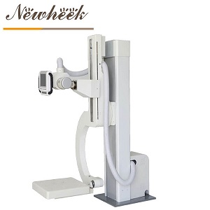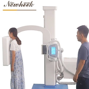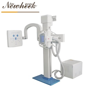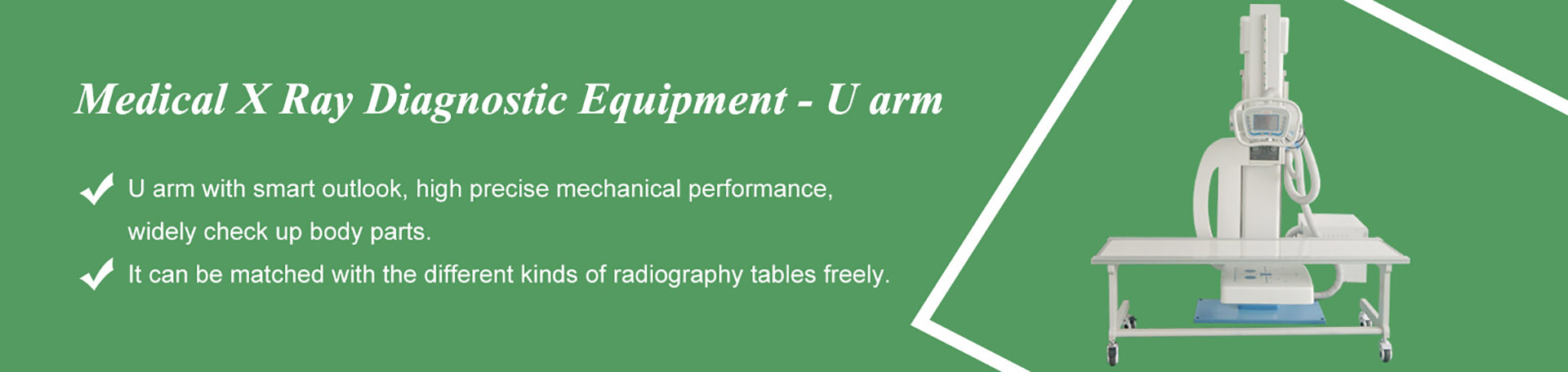

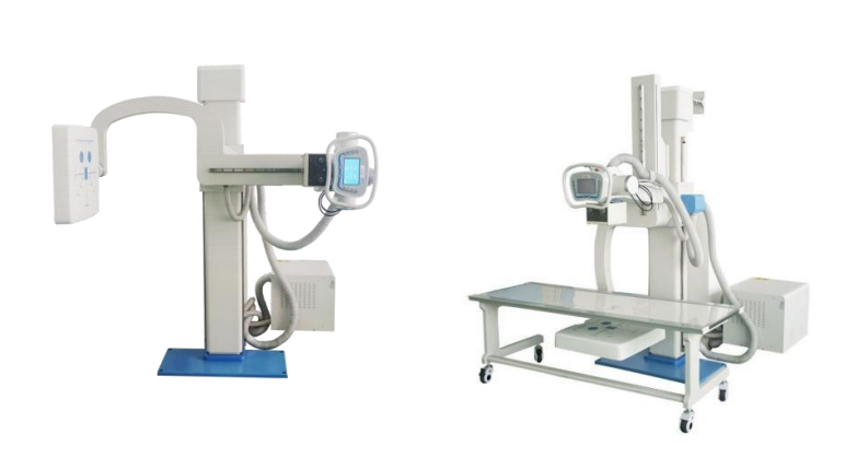
A U-arm X-ray system is a digital radiography machine whose X-ray tube and flat panel detector hang on opposite ends of one curved, column-mounted arm, so the pair moves around a stationary patient for standing, sitting and lying exams. Whether it can also handle lateral, oblique and cross-table projections is not a property of the name “U-arm” — it is a property of four components: arm rotation, bucky tilt, SID travel and the rigid tube-detector geometry. Some U-arm builds genuinely cannot angle; others are publicly documented doing exactly that. Here is how to tell them apart before you buy.

The claim is not invented — it is just applied too broadly. Under the “U-arm” label you will find two very different builds:
So the honest answer to “can a U-arm take angled views?” is: some can, some cannot, and the difference is spelled out on the specification sheet — not in the category name. That is good news for buyers, because specifications can be checked, compared and written into a purchase contract.
Before you shortlist any machine, dig four numbers out of the datasheet. They, more than anything else, determine which projections the room can actually produce:
| Component | What it decides | Commonly published range* |
|---|---|---|
| Arm rotation around the column | Whether the tube-detector pair can swing to the patient’s side for upright lateral and oblique work without turning the patient | Roughly −30° to +120° on some models, up to ±135° on others |
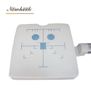
| Bucky / receptor tilt | Tilted-projection work (oblique spines, angled chest views) with the receptor kept square to the beam | Commonly around ±45° |
| SID travel, motorized | Correct source-to-image distance per exam — 180 cm chest technique down to shorter extremity distances | Roughly 100–180 cm |
| Single rigid arm carrying both ends | Alignment survives every angle: the tube and detector cannot drift apart while the arm sweeps | Geometry, not a number — verify at demo |
*Ranges vary significantly between models and manufacturers; treat these as the typical band found in public specifications, and always confirm against the specific model’s datasheet. For orientation, the vertical coverage figures commonly published alongside these ranges include a horizontal-beam height span of roughly 51–160 cm and a detector-center-to-floor span of roughly 62–170 cm.

One structural point deserves emphasis because it is what makes angled views reliable rather than merely possible: because both optical ends ride the same rigid arm, the tube-detector alignment that was verified at one angle still holds at every other angle. A build where tube and receptor are mounted on separate structures has to be re-aligned — or trusted — at each new projection.
Map your examination list onto the four components above and the “can it?” question turns into arithmetic:
If you want a deeper dive into which body parts a sickle arm covers, we have a dedicated walkthrough, as well as a focused look at chest films and lumbar spine work on a U-arm.
The second half of the familiar objection — that switching between chest and table positions takes longer than on other builds — deserves the same honest treatment. It is real, and it shows up when the schedule is heavy. Three countermeasures are standard practice in busy U-arm rooms:
Room geometry plays into this too — our guide to U-arm room requirements (space, ceiling height, floor loading, power and shielding) covers the layout side.
None of this makes the U-arm the best machine for everything, and it does not need to be. Imaging-industry explainers describe the division cleanly: straight columns excel at high-volume upright work; U-arms cover upright and supine in one compact room; C-arms belong to the operating theatre, where real-time fluoroscopic guidance — not static radiography — is the job. If you are weighing a U-arm against a C-arm, the real question is whether your list contains surgery, and our comparison of the two goes through it in detail.
The whole argument above compresses into one page of procurement language. Ask every supplier to answer these in writing:
A supplier who answers these fluently is selling you a machine they know. A supplier who answers “yes to everything” without numbers is selling you a category name. The same checklist works as an acceptance script after installation, and if you want the arm-handling side of the equation — how operators actually position patients to keep repeat rates down — that is covered in our positioning guide.
When you are ready to see how a specific configuration maps onto your examination list, send us the list. You can start from our multifunctional U-arm radiography system and take the geometry questions straight to the datasheet. For a component-by-component tour of the machine itself, our sickle arm structure explainer covers the motion ranges in depth.
Depends on the build. Multi-axis U-arms — where the tube and detector hang on one curved arm that rotates around the column and the bucky tilts — are publicly documented supporting AP/PA, lateral and oblique projections. Fixed straight-column builds generally are not. Check the four components: arm rotation, bucky tilt, SID travel and the rigid single-arm geometry.
Because they are describing the fixed straight-column build, or entry-level models close to it. The U-arm category spans a wide range of motion capability, which is exactly why the projection list belongs in your RFQ rather than in your assumptions.
A cross-table lateral is a horizontal-beam projection — the X-ray beam runs parallel to the floor while the receptor sits horizontally beside the patient — used in trauma and hip imaging where moving the patient is not an option. Support depends on the bucky or detector carriage accepting a horizontal orientation; ask the supplier to demonstrate it rather than confirm it verbally.
Industry comparisons note that chest-to-table changeover takes longer on U-arms than on some other builds, and it becomes noticeable at high volume. Motorized presets, batching exams by position and walkthrough operation are the standard countermeasures — and timing the changeover during your demo turns the objection into a number you can negotiate against.
Tell us your examination mix — chest, spine, extremities, trauma — and which angled views it depends on, and we will map it against the arm geometry before you commit. Fill in the inquiry form in our Contact Us module with your name, email, country and requirements, and our product manager will get back to you as soon as possible (within 24 hours on working days, Monday–Saturday) with a configuration recommendation. You can also reach us directly at admin@newheek.cn or WhatsApp +86 19062611512.
Tel: +86 19062611512
Email : admin@newheek.cn
Company : Weifang Newheek Electronic Technology Co., Ltd.