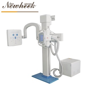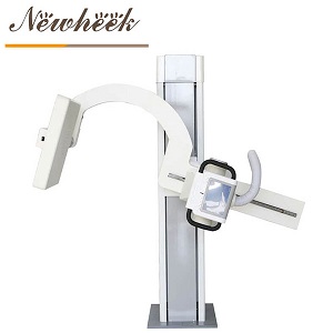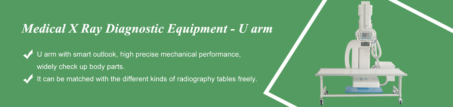

A sickle arm X-ray machine — also called a U-arm — is a digital radiography (DR) system that carries the X-ray tube and the flat panel detector on the two ends of one curved arm fixed to a floor-mounted column. Because both ends share the same rigid arm, the tube and detector stay aligned while the arm rotates, travels and sweeps around a stationary patient — one machine covers standing, sitting and lying examinations.
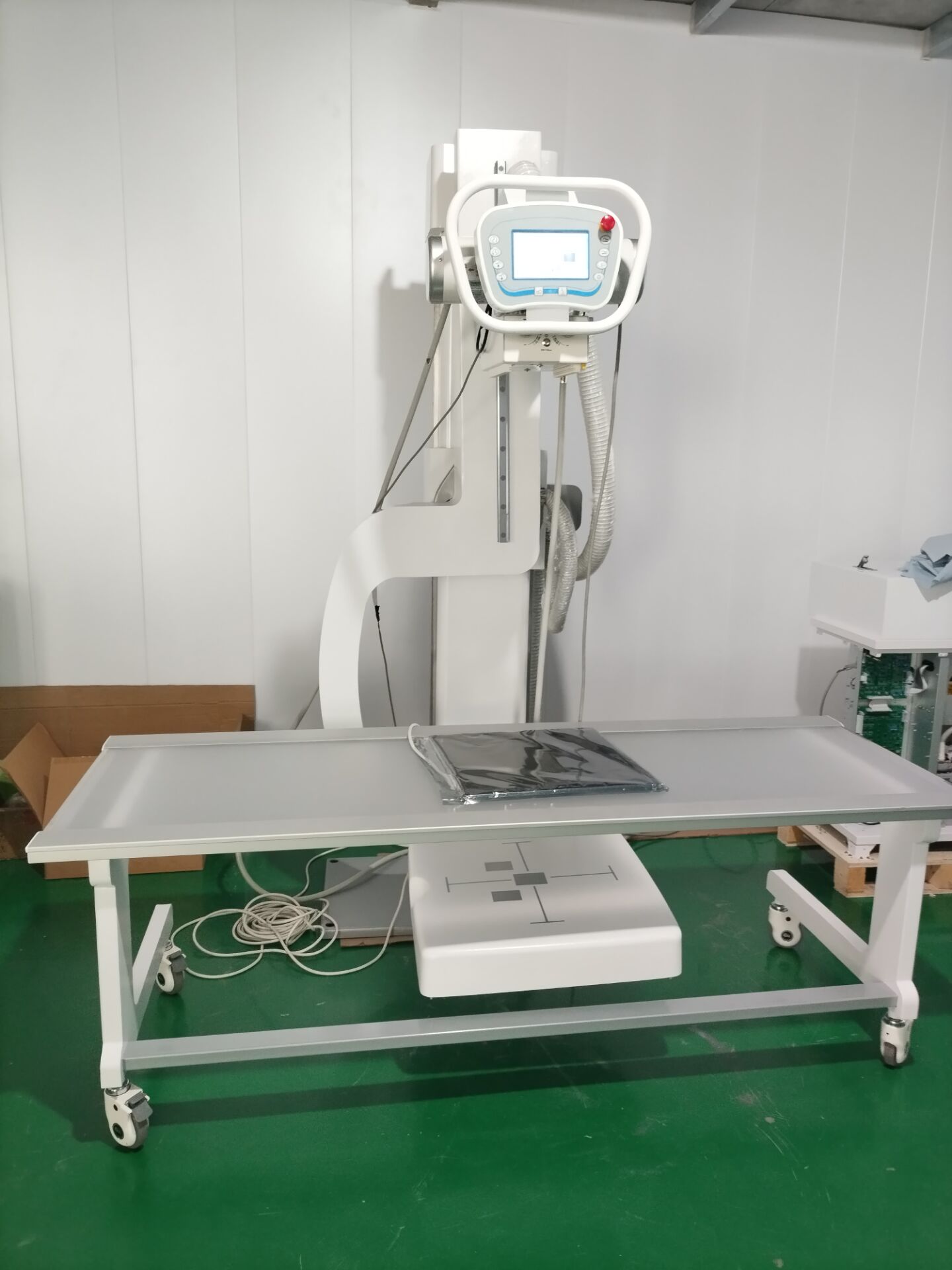
A complete sickle arm DR room in one frame: floor column, curved U-arm, radiographic table and hand-held console.
Before the specs, a naming map — because the same machine family is sold under four labels:
Practical buying advice: ignore the label and read the motion specification sheet — rotation ranges, vertical travel and SID. That is where the real differences between suppliers hide.
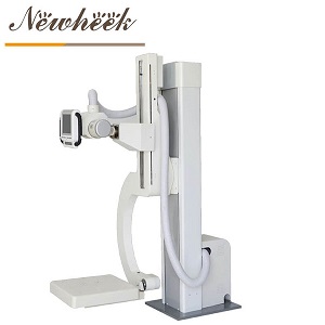
A sickle arm machine is an imaging chain of six subsystems mounted around one column: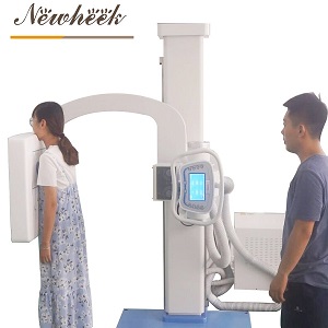
These are the published motion figures of the Newheek U200 / U300 sickle arm platform — the sheet a buyer should demand from any supplier:
| Motion | Specification | What it enables |
|---|---|---|
| Arm rotation (around the column) | −30° to +120° | PA, lateral and oblique projections without moving the patient |
| Tube rotation | ±90° | Independent beam angulation for tangential and angled views |
| Detector rotation | ±45° | Receptor stays square to the beam at every angle |
| Vertical travel of the arm | 1,200 mm stroke | From standing chest height down to a wheelchair or table-top position |
| Lowest arm position (to floor) | 480 mm | Extremity work with the patient seated or the limb resting on a stool |
| SID (tube-to-detector distance) | 1,000–1,800 mm, motorized | Upright chest at 1,800 mm, extremities at 1,000 mm — driven, not cranked |
| Cassette holder size | 620 × 580 mm | Accepts standard grid cassettes and digital panels |
The internet is full of “flexible, efficient, comfortable” advantage lists that say nothing. Here is the same case made with checkable evidence:
A sickle arm needs one floor column and wall space for the upright cassette holder — no ceiling rail, no reinforced ceiling. Market research on DR architectures (HDIN Research, DR Market Report 2026–2031) describes floor-mounted U-arm systems as minimizing the spatial envelope required for installation, often under 16 square meters, which is exactly why urgent-care clinics and community hospitals standardize on them. For the power side of the same calculation, see our power supply guide.
Because the tube and the detector hang on the same rigid arm, they cannot drift out of alignment. UK distributor Medigear summarizes the mechanism well: tube and detector held in fixed alignment on a single arm speeds positioning and reduces alignment errors — and every avoided alignment error is a retake that never happens, for the clinic and for the patient’s dose.
The −30° to +120° sweep plus the 1,200 mm vertical stroke means the machine moves around the patient, not the patient around the machine: upright chest at the wall bucket, seated shoulder at an angle, supine abdomen over the table. One installation replaces what used to need separate chest and general rooms.
The 1,000–1,800 mm motorized SID range is not cosmetic: 1,800 mm is the standard focus distance for upright chest imaging. Push-button SID also means switching between a chest exam and an extremity exam takes seconds, with no manual cranking between patients.
The console stores 3 preset positions — typically chest, table and extremity — so a repeated exam returns to position with one key. And because a heavy arm sweeping low could strike the floor or a table edge, rotation is blocked at low positions (anti-tip protection): the arm simply refuses the move. For high-throughput rooms with rotating staff, these two functions are what keep positioning consistent between operators.
The Newheek U-arm platform is built around a 50 kVA high-frequency generator (max 100 kV / 500 mA, tube current 25–630 mA), a 30.5 kW / 125 kV dual-focus tube (1.0 / 2.0 mm, 140 kJ anode heat capacity) and a manual collimator with a 440 × 440 mm maximum field. That headroom supports short exposure times at higher currents — which helps motion control on patients who cannot hold still, from young children to elderly patients.
To keep expectations accurate: a sickle arm is a digital radiography system — still images, one exposure at a time. It is not the tool for real-time fluoroscopic imaging around an operating table; that is the mobile C-arm’s job, a different rack with a different purpose. Some suppliers market “dynamic DR” platforms that add fluoroscopy-style acquisition on a U-arm base — if that is on your shortlist, compare the dynamic specification explicitly. (See also: U-arm vs C-arm, explained.)
Yes — two names for the same machine family: one curved arm carrying the tube and detector on opposite ends, fixed to a floor column. “UC arm” adds the combined independent rotation of arm, tube and detector.
Industry research puts floor-mounted U-arm rooms at often under 16 m². You need floor space for the 2,200 mm column, wall space for the upright cassette holder, and clearance for the arm sweep — usually less equipment room than any ceiling-suspended alternative.
Yes. The motorized SID extends to 1,800 mm — the standard distance for upright chest radiography — and the arm rotates so the tube-and-detector pair faces the wall-bucket position for PA and lateral chest work.
To 480 mm from the floor. That low position brings the detector down to wheelchair patients and to limbs resting at seat height, without transferring the patient onto a table.
A standard sickle arm is a static digital radiography system — still exposures only. Real-time fluoroscopy belongs to the C-arm category; some “dynamic DR” platforms add it on a U-arm base, which should be compared by its dynamic specification, not by the U-arm label.
Newheek ships within 15 workdays after payment receipt, and third-party inspection is supported before delivery.
Tell us your room size, power supply and main examination mix — simply fill in the inquiry form in our Contact Us module with your name, email, country and requirements, and our product manager will get back to you as soon as possible (within 24 hours on working days, Monday–Saturday) with a configuration recommendation and a formal quotation. You can also reach us directly at admin@newheek.cn or WhatsApp +86 19062611512.
Tel: +86 19062611512
Email : admin@newheek.cn
Company : Weifang Newheek Electronic Technology Co., Ltd.