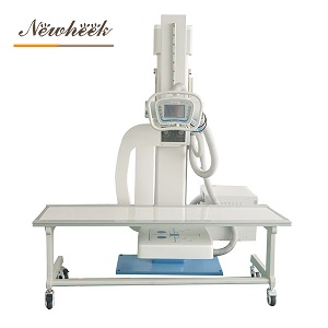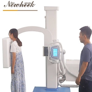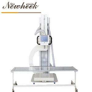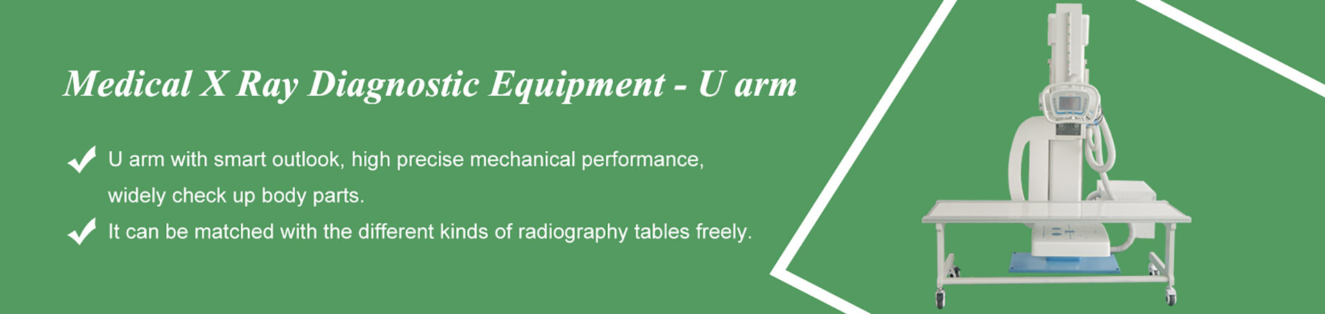

A UC arm X-ray machine is a digital radiography (DR) system whose X-ray tube and flat panel detector are mounted on the two opposite ends of a U-shaped arm fixed to a floor-mounted column. The tube and detector stay perfectly aligned while the whole arm rotates and travels vertically around a stationary patient — so one machine covers standing, sitting and lying positions without re-positioning the patient. In common industry usage, “UC arm” refers to a U-arm that also offers combined rotation of the arm, the tube and the detector — a “U + C” motion range that a basic single-pivot U-arm does not provide.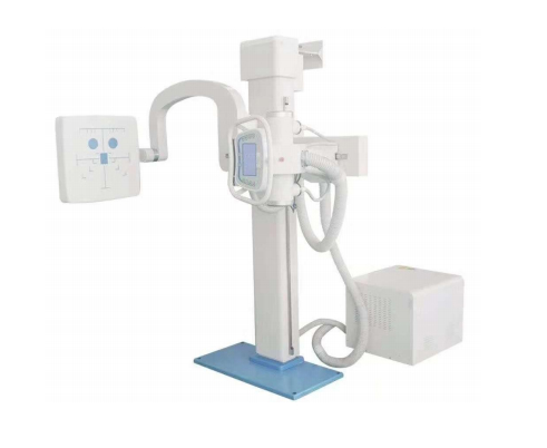
1. Where the Names Come From — and Why They Get Mixed Up
The naming in this product category is genuinely confusing, so let’s clear it up first:
U-arm (sickle arm) — the geometry. The tube head and the detector sit at the two open ends of a rigid U- (or sickle-) shaped arm. Because both ends are fixed to one arm, tube and detector are always centered on each other, no matter where the arm moves.
UC arm — the motion capability. “UC” is widely used in the industry for a U-arm platform whose arm, X-ray tube and flat panel detector can each rotate independently, so the beam angle can be changed the way a C-arm does, while keeping the U-arm’s stability and detector alignment. Think of it as “U-shaped arm, C-style angulation.”
U-arm DR / UC-arm DR — the same machines once a flat panel detector and a digital workstation are added. In practice, “DR” simply states that the system captures images digitally instead of using film.
A practical note for buyers: many suppliers use “U-arm” and “UC arm” interchangeably in their marketing pages. The reliable way to compare machines is not the name — it is the motion specification sheet: rotation ranges, SID travel and preset positions. More on that below.
2. The Anatomy of a UC Arm DR System: Six Subsystems
A UC arm machine is not one device — it is an imaging chain of six subsystems working together: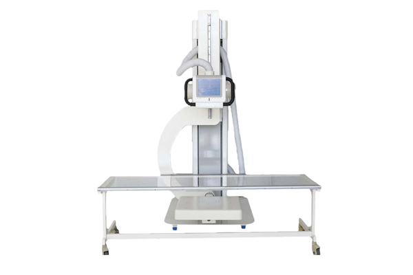
| Subsystem | What it does | Typical spec on a Newheek UC arm |
|---|---|---|
| X-ray HV generator | Feeds controlled power to the tube | Max output 50 kVA (100 kV / 500 mA), 25–630 mA |
| X-ray tube | Produces the beam | 30.5 kW / 125 kV, dual focus 1.0 / 2.0 mm, 140 kJ anode heat capacity |
| Collimator | Limits the field, shows the light field | Manual, field up to 440 × 440 mm at SID 100 cm |
| UC arm rack | Carries and moves tube + detector | Vertical travel 1,200 mm; lowest arm position 480 mm from floor |
| Flat panel detector | Converts X-rays into digital images | a-Si, 14″ × 17″, 150 μm pixel pitch, 3.3 lp/mm (see also our notes on flat panel detectors) |
| Image workstation | Acquisition, processing, print, DICOM transfer | Full image in approx. 5 s after exposure |
Both types share the same U-shaped geometry. The difference shows up in how many ways the system can rotate. Here is what a full UC-arm motion set looks like, using the published figures of the Newheek U200 / U300 platform:
| Motion | Basic U-arm | UC arm (U200 / U300) | Why it matters |
|---|---|---|---|
| Arm rotation (around the column) | Often a single sweep | −30° to +120° | Covers PA, lateral and oblique projections on one machine |
| Detector rotation | Fixed to the arm | −45° to +45° | Keeps the receptor square to the beam at angled positions |
| Tube rotation | Fixed to the arm | ±90° | Tube angle can be set independently for tangential views |
| SID travel (tube-to-detector distance) | Manual, limited | 1,000–1,800 mm, motorized | Chest work at 1,800 mm, extremities at 1,000 mm — no repositioning |
| Vertical travel | Limited | 1,200 mm stroke (600–1,550 mm working range) | From a wheelchair patient down to a table-top extremity |
| Preset positions & anti-collision | Rare | 3 presets + rotate-protection | One key returns the arm to a saved position; rotation is blocked at low positions so the arm cannot strike the floor or table |
In short: a basic U-arm moves; a UC arm positions. The independent rotations and motorized SID are what let one room handle chest, spine, abdomen, skull and extremity work with a single patient placement.patient placement.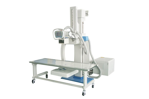
Where does the UC arm sit among the other DR rack types?
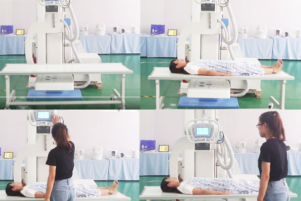
They describe the same U-shaped family, but “UC” emphasizes the combined rotation of arm, tube and detector. When comparing suppliers, judge by the motion specifications, not the label.
Different jobs. A mobile C-arm does intra-operative fluoroscopy around an operating table; a UC arm does high-volume digital radiography in a dedicated room, with much better mechanical stability for standing radiography.
Yes — the motorized SID extends to 1,800 mm, which is the standard focus distance for upright chest imaging, and the wall-bucket position supports PA and lateral chest work.
Two: Model U200 and Model U300, built on the same 50 kVA imaging chain. The U300 configuration is matched with X-ray radiography tables for supine examinations.
Three-phase 380 V ±10% power with ≥55 kVA capacity, grounding resistance ≤2 Ω, and floor space for the 2,200 mm column plus patient approach. Installation specifics are usually confirmed with the supplier before shipment.
Newheek ships within 15 workdays after payment receipt, with third-party inspection supported before delivery.
If you are deciding between a U-arm and a UC arm for your room, tell us the space, power supply and main examinations — simply fill in the inquiry form in our Contact Us module with your name, email, country and requirements, and our product manager will get back to you as soon as possible (within 24 hours on working days, Monday–Saturday) with a configuration recommendation and a formal quotation. You can also reach us directly at admin@newheek.cn or WhatsApp +86 17616362240.
Tel: +86 19062611512
Email : admin@newheek.cn
Company : Weifang Newheek Electronic Technology Co., Ltd.