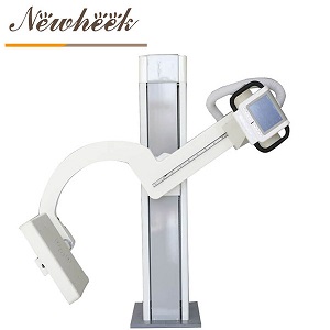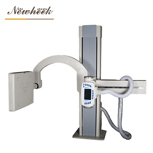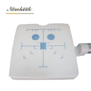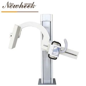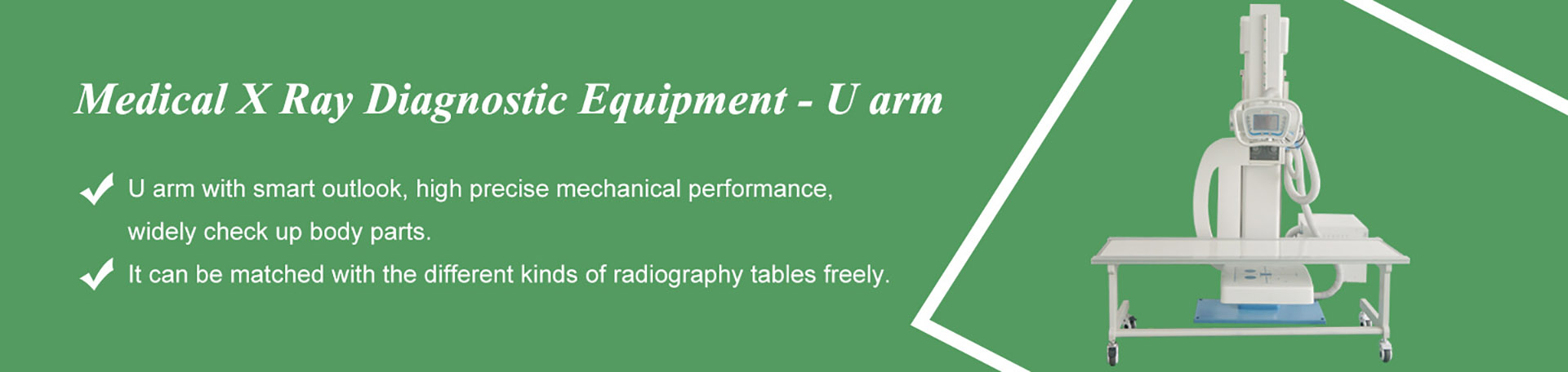

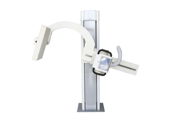
A sickle arm (also called a U-arm) is an X-ray machine built around one simple mechanical idea: the X-ray tube and the flat panel detector are locked onto the same rigid arm and move together, around the same axis, as one unit. That single design fact is where every real advantage of this machine comes from — and it is also the fastest way to decide whether the machine fits your room.
So before we list use cases, here is the honest one-line version: a sickle arm is at its best wherever you need to reposition the beam quickly around a patient who stays put — standing chest exams, orthopedic follow-ups, screening lines, small clinics, and uncooperative veterinary patients. It is the wrong tool wherever you need a large moving picture, or angles that reach around the patient. The rest of this article unpacks both halves of that sentence.
On a classic radiography room with a floor tube stand, the tube and the wall stand are two separate devices. Every position change means moving two things and hoping they stay aligned. On a sickle arm, the tube and the detector cannot drift apart, because they are physically one assembly. When the arm rotates, the beam and the receptor rotate with it, already centered on each other.

Everything in the list below is a consequence of that fact. Keep it in mind, because it is also the checklist you should run through when a salesperson shows you the machine: every claimed advantage should trace back to something you can watch the arm do.
Watch what happens in an orthopedic follow-up. The patient arrives in a cast, or limping, or holding a brace against their ribs. On a two-device setup, getting a lateral view of an injured ankle can turn into a negotiation: stand here, turn your foot, hold still, now more. On a sickle arm, the patient steps into position once, and the arm does the traveling — swinging from front-facing to side-facing while the patient barely moves.
This matters far beyond orthopedics. Elderly screening patients, post-operative visits, anyone in pain: every position change you ask of them is time, discomfort, and a chance for motion blur. The design moves the complexity away from the person on the receiving end.
Because the tube-and-detector pair rotates as a unit, a patient who is already standing against the arm can get a frontal view and then a lateral view without stepping off, sitting down, or re-gripping the handles. In a physical-exam line that processes one person every few minutes, this is the difference between a smooth conveyor and a waiting room full of people wondering what is taking so long.
The same property pays off in orthopedic clinics that want weight-bearing views: the patient stands, the arm swings, and you compare front and side projections taken minutes apart with the patient in essentially the same stance.
A sickle arm is a column-based machine. It does not need a full floating tube stand, a wide floor track, or the clearance that a table-plus-tube layout demands. In practice this is why the design shows up so often in single-room clinics, converted office spaces, and screening units where the X-ray room shares its square meters with everything else the facility does.
One caution from the field: the machine being compact does not mean any room works. Ceiling height, floor condition and power supply still decide whether installation is straightforward. If you are still choosing or preparing the room itself, read the room requirements guide first and save yourself a return visit from the installers. (We cover this in detail in our room requirements article.)
Here is an advantage almost nobody puts on a brochure, and it is the one experienced radiographers care about most. Because the arm returns to the same geometry reliably — same height, same angle, same source-to-image relationship — a fracture photographed at week one and again at week six comes out with the projection matching. The doctor is comparing the bone, not comparing two different camera angles.
For screening programs, occupational health checks and orthopedic follow-up series, this repeatability is quietly worth more than any single image quality claim. Ask any provider to demonstrate it: take a phantom or a volunteer in one position, move the arm away, bring it back, and compare. A well-built arm comes home.
On most modern units, the arm’s main movements — height, arm angle, and tracking — are driven from controls that do not require the operator to stand beside the tube at the moment of exposure. Combined with the standard controlled-area practices every X-ray room already follows, this keeps routine workflow simple: position the patient, step back, expose, swing to the next projection.
We are deliberately not listing numeric specs here. Distances and dose numbers belong in a quotation and a protection plan, not in a generic article — ask your supplier to walk through both with your actual room dimensions.
| Setting | What the workload looks like | Which design point carries it | What to verify on a demo |
|---|---|---|---|
| Orthopedic clinic | Follow-ups, post-reduction checks, cast series, standing weight-bearing views | Move-the-machine-not-the-patient; repeatable geometry for comparable series | Swing from AP to lateral with a volunteer holding still; check how precisely the arm returns to a marked position |
| Physical exam & screening center | Long lines of standing chest exams, one person every few minutes | One stance, multiple projections; fast repositioning between patients | Time a full cycle: patient in, front view, side view, patient out. Then repeat with the next “patient” |
| Small or single-room clinic | Chest, limbs and spine in one shared room, tight floor plan | Column design hugs the wall; no separate tube stand footprint | Bring your room sketch. Confirm column footprint, door width and turning space against your actual plan |
| Emergency & urgent care | Fast answers for walking wounded; seated patients; follow-up imaging | Quick repositioning around a stationary patient | Discuss your non-standing patient share. For wheelchair and seated workflows, see our dedicated article on upright workflows for patients who can’t stand |
| Veterinary practice | Animals will not hold a pose; sedation is costly; throughput matters | Fast positioning around an uncooperative subject; exposures from a distance | Ask how the arm behaves at low table heights and what positioning aids exist for animal work |
| Occupational & follow-up programs | Same patients, same views, months apart; results must be comparable | Geometry that comes back to the same position visit after visit | Ask how positions are recorded and reproduced, and whether the console saves preset positions per protocol |
Two of these rooms deserve an honest footnote. For emergency work, a sickle arm handles the walking wounded well, but patients who genuinely cannot be moved belong to mobile radiography, and seated workflows have their own logic — we wrote a separate guide on chest X-ray workflows for patients who cannot stand. For veterinary work, large animals bring their own constraints; we also supply machines configured for cattle and pig lung screening, and the positioning habits there differ from human clinical work.
An article that only lists advantages is an advertisement, not a guide. So here are the jobs this design should not be given:
If your reading of the six-room table left you unsure which side of this line your workload falls on, that is normal — send us the case mix and we will tell you plainly whether the arm fits, including the cases where the honest answer is that you need a different machine and we would rather say so than sell you the wrong one.
Every advantage above is verifiable in a fifteen-minute demonstration. Do not let the demo become a slideshow; insist on hands-on:
Bring your floor plan and your case mix to the demo. A supplier who answers both without dodging is a supplier whose other claims you can start trusting.
Yes. The names describe the same design from different angles: the arm that holds tube and detector together is shaped like a “U”, and its silhouette also resembles a sickle. You will see both terms, and “UC arm” as well, used interchangeably across the industry. We unpack the naming in our plain-language guide to the sickle arm structure.

Yes. The arm covers a standing patient against the column, and with a photography table in front of it, it covers supine and reclined positions as well. The pair of a sickle arm and a floating table is the most common small-room configuration we ship. If you are comparing table options, our bucky stand guide covers the upright piece separately.
Compact footprint helps, but shielding and power depend on your local regulations and the machine’s output, not on the arm’s shape. Do not sign a lease before reading the room requirements guide — retrofitting a room that failed the survey is the most expensive mistake in this purchase.
Increasingly, yes — the fast repositioning and distance operation suit animal work well, and we ship veterinary configurations. The differences from human clinical use are mostly in positioning aids, table height and cleaning expectations, so tell your supplier the species mix up front.
Three things: your room dimensions including ceiling height, your expected daily case mix by body part, and whether your patients can mostly stand. With those three answers a serious supplier can tell you within one email whether a sickle arm is the right category at all — and which questions you should be asking next.
If you are weighing a sickle arm against other designs for a specific room, send us the floor plan and case mix — we will tell you where the design fits and where it does not. Reach the factory team directly:
You can also browse the U-arm digital X-ray machine product page for configuration details before you write.
Tel: +86 19062611512
Email : admin@newheek.cn
Company : Weifang Newheek Electronic Technology Co., Ltd.