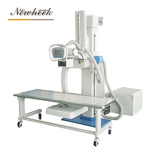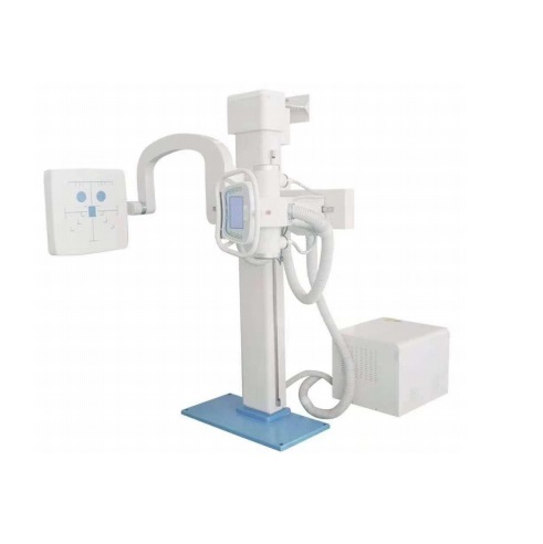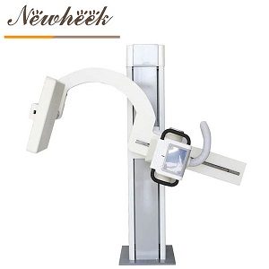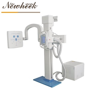

Quick answer: the collimator is the unglamorous box sitting directly under the X-ray tube — the one with the light and the two knobs (or buttons). It does two jobs: it shows the radiographer exactly where the beam will land, using a light field, and it crops the beam down to just the body part being examined. That second job is why this small part quietly shapes everything buyers actually care about: a well-collimated image is sharper because less scatter fogges it, the dose is lower because tissue outside the frame never receives radiation at all, and there are fewer repeats because the anatomy that matters is actually inside the picture. This article explains the part in plain language, and lists what to check before you buy — no model numbers required.
Scope note: this article deliberately names no collimator models and quotes no certification marks. Which collimator pairs with which machine is a question with a written answer from your supplier — the checklist below tells you what to ask for and why.

Picture two chest images from two rooms. One is grayish, low-contrast, the kind of image a radiologist squints at. The other is crisp — bones bright, lung fields clear. The manager of the first room has already budgeted for a better detector. And yet the difference between the two rooms is sometimes not the detector at all. It is how much of the beam was cropped before it ever reached the patient.
Here is the mechanism, minus the physics degree. When X-rays pass through a body, some of them scatter — they bounce off tissue and travel onward in random directions, like light through fog. Every square centimeter of tissue inside the beam adds fog to the image. Crop the beam to just the chest, and you get the fog from the chest. Leave the beam wide open and half the abdomen inside it, and the abdomen contributes fog too — for no diagnostic benefit whatsoever.
The part that does the cropping is the collimator. It has no screen, no software, no marketing story. It is a frame with adjustable blades and a small light. And it is quietly one of the highest-leverage components in the room.

Strip away the terminology and a collimator does two things, both easy to picture:
That is the whole device. Which raises an obvious question: if it is that simple, why does it deserve an article? Because the simplicity is exactly where it gets underestimated — both in daily habits and in purchase decisions.
Collimation is the rare habit that pays out in three currencies simultaneously:
Three currencies, one pair of blades. It is also why the collimator is one of the few components whose importance grows with patient anxiety: a visible light field reassures the person on the table that only the necessary area is involved. Small feature, outsized trust.

Online discussions about collimators tend to collapse into one question — “which model fits my machine?” — which is exactly the wrong place for a buyer to start, because the answer is a supplier lookup, not a decision. The decisions live somewhere else. Here is the checklist worth thinking through:
Notice that four of the five items are about behavior over time — alignment honesty, workflow fit, repeatability. That is consistent with how the rest of a good machine purchase works: the column layout decision gets the attention, but the small parts decide whether the room feels tight or sloppy in daily use. If you are still weighing layouts, our comparison of the three column layouts pairs well with this checklist.
Buying a good collimator is half the value; the other half is a habit that costs two minutes to learn and lasts a career: crop before you expose, every time, on purpose. Open the light field, shape it to the anatomy, check the edges, then expose. Not “usually.” Every time — because the exposure that never happens is the only dose reduction with no trade-off at all.
Two populations make the habit non-negotiable. Children, whose smaller bodies mean a loosely cropped beam covers proportionally more of them — our pediatric and neonatal U-arm guide builds the same principle into positioning from the start. And repeat patients, whose cumulative exposures are the sum of every habit in the room. Collimation is where a room’s quiet discipline — or the lack of it — becomes visible in the films years later.
The good news: this is one of the few “upgrades” in radiography that costs nothing. The box is already on the machine. The only purchase is the habit.
Two jobs: it projects a light field showing exactly where the beam will land, and it crops the beam with adjustable blades so only the anatomy of interest is exposed. It is the frame around the picture — and the reason a framed image is sharper and lower-dose than a wide-open one.
Yes, and in the most direct way possible: tissue outside the cropped field receives essentially no radiation, because the beam is blocked at the source. No processing trick or detector upgrade reduces exposure for anatomy that was never in the beam. It is the first dose-management habit in radiography for a reason.
Less tissue in the beam means less scattered radiation fogging the detector, so the signal is cleaner and contrast is higher. A gray, washed-out image is often a wide-open beam story rather than a “we need better equipment” story.
Depends on the room’s workflow. Manual blades are mechanically simple and suit lower-throughput rooms; electric blades are driven from the console and save steps in busy rooms. The honest comparison runs through your exam volume and who operates the room — ask the supplier to walk both against your workflow.
Four things, in writing: which collimator ships with the machine, whether manual and electric options exist, how light-field alignment is checked and corrected over time, and which regulatory documentation is provided for your region. A supplier who answers these easily has thought the machine through past the headline specs.
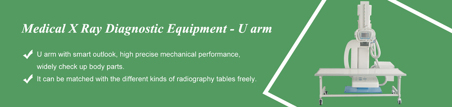
When you specify a U-arm system, the collimator questions above deserve real answers, not brochure nods. Tell us your exam list and workflow; we will reply with which configuration we would recommend and the documentation that accompanies it — in plain language, for your file.
Email: admin@newheek.cn | WhatsApp / Phone: +86 19062611512
Tel: +86 19062611512
Email : admin@newheek.cn
Company : Weifang Newheek Electronic Technology Co., Ltd.