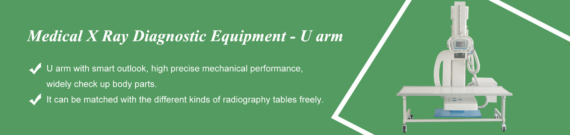

a U-arm system can serve pediatric and neonatal work well — but only if three things are true. First, the room was selected with children in mind: low detector travel, fine positioning control, short exposure capability and pediatric technique presets verified in writing with the vendor. Second, dose is managed as a practice, not a brochure claim: pediatric protocols, tight collimation, and a repeat-exposure rate the department actually measures. Third, immobilization is planned as a workflow — positioning aids, carer involvement defined in advance, and shielding policy aligned with current international guidance and local regulation. This article covers all three from the machine-and-workflow side: what to check before you buy, what to do differently in the room, and what to put in the RFQ so the answers come back in writing.
Scope note: this article addresses pediatric imaging from the equipment-selection and workflow perspective of a U-arm radiographic room. Detector-side topics — panel sizes, scintillators and pediatric detector presets — are covered in depth in our sister article on pediatric radiography and the detector. No absolute dose figures are given here by design: dose limits and protocol values belong to your medical physicist and local regulation. Whether any specific configuration supports a stated feature must be confirmed with the manufacturer before purchase.
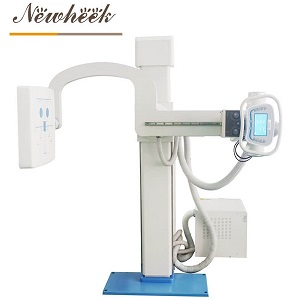
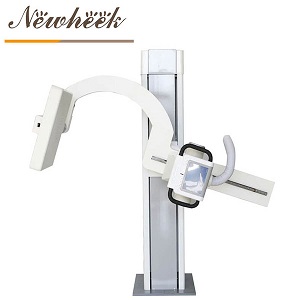
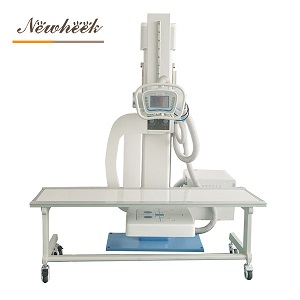
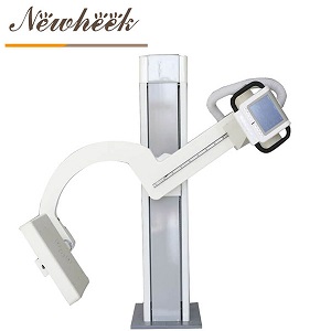
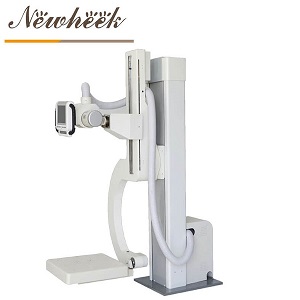
A U-arm carries both the X-ray tube and the detector on one rotating arm, which is precisely the property pediatric rooms need: the geometry can come to the child, instead of the child being moved to the geometry. A sleeping infant on the table can be imaged with the arm rotated to a horizontal, over-table geometry. A toddler who will stand but not lie down can be imaged upright at the wall stand — even a distressed, wiggling one — with the arm tracking their height. An infant held by a carer can be imaged in whatever position keeps everyone’s hands out of the beam.
That flexibility is the honest case for a U-arm in pediatric service. But it only materializes if the arm’s movement characteristics match small, delicate positioning work:
None of these show up in a hero photograph of the machine. All of them belong in the RFQ.
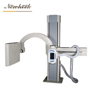
Manufacturers sell most U-arm rooms as general-purpose. That is not a defect — but it means pediatric capability is something you must verify, not assume. The differences concentrate in four areas:
| Area | General-room default | What pediatric service needs you to verify |
|---|---|---|
| Positioning range | Adult standing chest and table heights | Minimum bucky/detector height; arm geometry at low positions; whether the tube head can reach over-table horizontal geometry for supine infants |
| Presets | Adult protocols | Whether pediatric technique presets exist, how many age/size groups they cover, and whether they can be edited by the department’s physicist |
| Exposure control | AEC chambers tuned for adults | AEC behavior at low dose pediatric settings; shortest achievable exposure pulse; whether pediatric AEC chambers can be selected independently |
| Collimation | Manual collimator | How tightly the field can be collimated for small anatomy; whether a light field is clearly visible for fast, accurate fielding on a moving patient |
Two of these deserve emphasis because they are the ones buyers discover after installation:
Presets are editable or they are decorative. A factory preset labeled “pediatric” that the department cannot modify will drift out of alignment with the physicist’s protocol within months. The RFQ question is not “do you have pediatric presets?” but “can our medical physicist edit exposure factors per age group, and is that edit logged?”
AEC designed for adults can misfire on infants. Chambers sized and calibrated for adult anatomy can produce inappropriate exposures when a small body covers only part of the chamber. Ask the vendor to describe, in writing, how their AEC is intended to be used for pediatric patients — dedicated pediatric chambers, manual technique, or something else — and to demonstrate it during acceptance testing with a pediatric phantom.
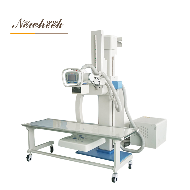
Radiation protection for children follows one organizing principle — ALARA: exposure kept As Low As Reasonably Achievable, recognizing that children have longer lifespans ahead of them for any dose effect to matter in, and that their smaller bodies need less radiation to make an image in the first place. Public guidance from bodies such as ICRP and the Image Gently alliance has shaped how well-run pediatric departments operate. The equipment’s role in that principle is real but bounded — roughly three layers:
We have covered the repeat-exposure problem for U-arm rooms generally in a dedicated article; for pediatric work, treat that content as dose management, not just efficiency. And be appropriately skeptical of any vendor who answers a pediatric dose question with an adjective (“low-dose”, “child-friendly”) instead of a mechanism (pulse behavior, preset editability, AEC design) and an offer to demonstrate it.
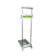
A calm, brief, well-planned examination is the best immobilization device that exists. Everything that lengthens the room visit — hunting for accessories, negotiating with a frightened toddler, re-centering by trial and error — raises motion risk and repeat risk together. Well-run pediatric departments plan immobilization on three levels:
One caution cuts the other way: restraint is a skill with a regulatory frame. How patients may be held, by whom, and with what documentation is defined by your institution and jurisdiction. Machine capability never substitutes for that framework — it only determines whether the framework can be executed quickly.
True neonatal imaging (in the NICU, on incubator-mounted or dedicated equipment) is its own discipline, and a general radiographic room will not replace it. But a U-arm room will see neonates and ex-premature infants — as outpatients, as follow-ups, and in facilities that share one room across ages. Three implications:
Pediatric claims are cheap in brochures and verifiable in documents. Put these eight questions to every bidder:
The vendor who answers these fluently has thought about pediatric work; the vendor who answers “the machine is universal” has not. Both answers are useful.
For outpatient and follow-up neonatal examinations, yes — provided the detector reaches table height, the collimator fields tightly, and exposure pulses are short. Dedicated NICU imaging (incubator-side, mobile) is a different service with different equipment and should not be improvised in a general room. Ask your vendor to state the room’s minimum patient size capability in writing, and validate it at acceptance with a phantom.
Current international consensus has moved away from routine gonadal/fetal shielding for most radiographic examinations, because precise collimation makes the benefit negligible and shields can interfere with exposure control and cause retakes — which are themselves dose. However, local regulation and institutional policy govern what your department must do. Follow your medical physicist’s written protocol; this article intentionally gives no numbers or absolute rules.
Not inherently — but the U-arm’s single rotating arm carrying both tube and detector makes pediatric repositioning fast: the geometry rotates around the child instead of the child moving between devices. For low-to-mid volume rooms, that workflow advantage is real. High-throughput pediatric hospitals may still prefer ceiling systems for other reasons. Match the equipment to the workload, and verify the pediatric-specific items of Section 2 either way.
Measure and attack the repeat-exposure rate. Retakes are the largest preventable dose multiplier in routine radiography, and their causes — motion, mis-collimation, wrong technique — are workflow failures, not equipment failures. A room with pediatric presets, editable by the physicist, plus a monthly repeat review will outperform a “low-dose” badge on a brochure every time.
Specifying a U-arm room that must handle patients of every size? Send us your pediatric requirements — we manufacture U-arm systems and will answer the eight questions of Section 6 for our own equipment in writing. You can also reach us directly at admin@newheek.cn or +86 19062611512.
Tel: +86 19062611512
Email : admin@newheek.cn
Company : Weifang Newheek Electronic Technology Co., Ltd.