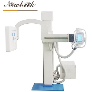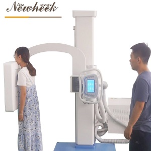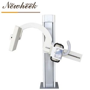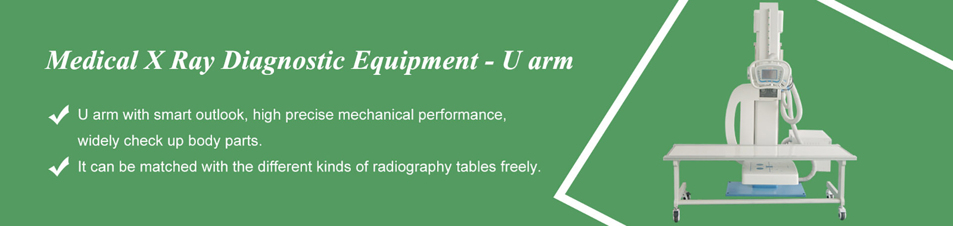

Quick answer: the reason a U-arm helps with difficult patients is not a special accessory — it is the geometry. The tube and the detector ride on the same arm and stay aligned while the arm rotates around the patient. For someone who is frail, in pain, or hard to transfer, that single fact changes the exam: the patient is positioned once, and the machine does the traveling. This article breaks down what that means in practice for three patient groups — elderly, bariatric, and limited-mobility — with the workflows, the boundaries, and the questions worth asking before the exam starts.
Contact: Email admin@newheek.cn · Phone/WhatsApp: +86 19062611512
A note on scope: we already have a dedicated article on upright chest imaging for patients who cannot stand. This article deliberately does not repeat the chest topic. Here the focus is everything around it — full-body positioning workflows on and around the U-arm’s table and wall-stand, for patients who make positioning genuinely difficult.

Three groups come up again and again in real rooms, and each one stresses a different part of the workflow:
Different as they are, all three groups reward the same design philosophy: minimize patient movement, maximize machine movement. That is exactly the trade a U-arm is built for.
On a conventional setup, changing the projection angle often means asking the patient to turn, lean, or rotate — instructions that a frail or painful body may not be able to follow. On a U-arm, the tube and detector are locked into opposite ends of the same C-shaped arm. Rotate the arm and the projection angle changes while the relationship between tube and detector stays fixed.
Translated into a difficult-patient workflow, this means:
For the projections that genuinely require different geometry — lateral decubitus, cross-table angles — the honest answer is that planning matters, and we cover those limits explicitly further down.
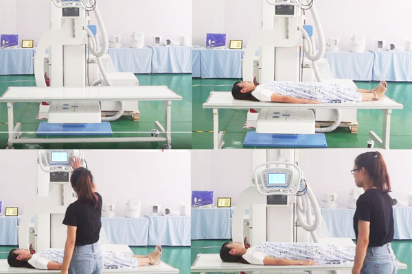
With elderly patients, the workflow goal is to make the exam short, boring, and safe. Boring is the compliment.
The ergonomics payoff is not only for the patient. Staff who would otherwise lift, steady, and re-lift a frail patient are the same staff who get injured doing it — the injury-data argument we made for motorized bucky stands in our motorized-vs-manual analysis applies with even more force to a frail population.
For bariatric patients, positioning problems start before any pose is attempted — they start with the room. A machine that sits too close to a wall, a table approach path cluttered with equipment, or a turning circle that does not fit a wider wheelchair all make the exam harder before it begins. That is why we tell buyers to audit the room against the machine before delivery: clearances, door widths, and approach paths are positioning equipment, even though nobody calls them that. Our room-requirements checklist walks through that audit.
During the exam itself, three habits carry most of the value:
That last point is worth underlining: we deliberately do not quote a universal “bariatric capacity” figure here, because real capacity depends on the specific table, stand, and configuration in your room. The defensible workflow is to know your population and verify the spec chain, in writing, per component.
For wheelchair users, stretcher patients, and anyone with restricted range of motion, the positioning rule is the one this whole article keeps returning to: do not ask the body to do what the machine can do.
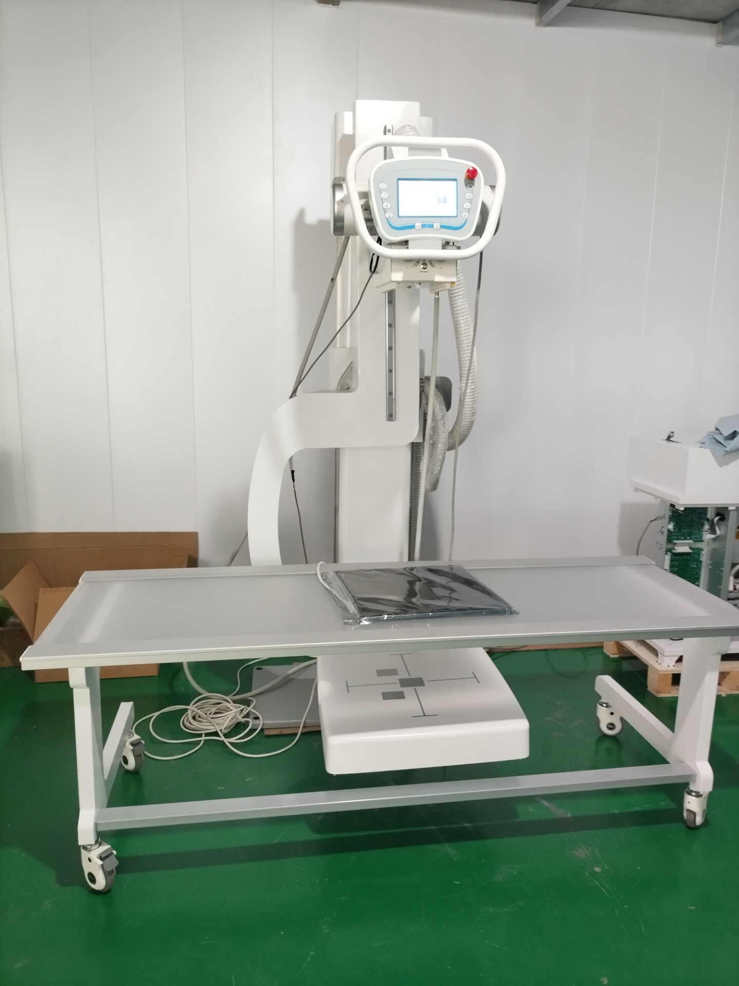
Before: know your views; sequence them by patient position; clear the transfer path; decide in advance whether the exam happens in the chair, on the table, or both — and only both if the extra transfer buys real information.
During: narrate the plan; batch same-position views; let the arm rotate instead of the body; give the patient a job they can do (“hold this, breathe normally”) instead of one they cannot.
After: help with the first movement off the table; note which positions worked and which did not, so the next difficult exam starts from experience instead of from zero.
Two boundaries matter, and pretending they do not exist helps nobody:
Frequently yes, depending on the views ordered. Supine and seated geometries cover a wide range of whole-body work, and the arm’s rotation supplies most projection changes. Whether your specific exam list fits a no-standing protocol is worth rehearsing with your team using your most common orders.
The arm’s patient-side advantages — machine moves instead of patient, fewer transfers, flexible geometry — apply fully. But capacity and clearance are component-specific: verify the table, stand and accessory specifications with your supplier in writing, and audit the room’s clearances before the patient’s first visit. We do not quote a universal figure because none is honest.
Sometimes — seated extremity and certain seated views are achievable when the chair’s frame stays out of the field and the arm reaches the required geometry. When either condition fails, a planned transfer onto the table is usually safer than an awkward pose held inside a wheelchair.
Improvising the sequence in the room. Difficult-patient exams reward planning — views ordered by position, transfers batched, and the arm’s rotation exploited — and punish whoever tries to design the workflow while the patient is already lying on the table in pain.
The core enabling feature is the arm geometry itself, not an add-on. Support accessories like positioning aids or mattress options exist and are worth discussing with your supplier based on your patient mix — but the transfer-reduction logic in this article works on the standard configuration.
Difficult patients do not need a different radiography machine as often as they need a different workflow around the one you have. The U-arm’s patient-stays, machine-moves geometry is purpose-built for exactly this population: elderly patients get shorter, safer exams; bariatric patients get room to be examined with dignity; limited-mobility patients get imaged where their bodies actually are. Plan the sequence, batch the transfers, rotate the arm — and if you are equipping a room where difficult patients are the norm rather than the exception, browse our U-arm digital X-ray machines and tell us about your patient mix so we can help you match the configuration to it.
Contact: Email admin@newheek.cn · Phone/WhatsApp: +86 19062611512
Tel: +86 19062611512
Email : admin@newheek.cn
Company : Weifang Newheek Electronic Technology Co., Ltd.