
Quick answer: orthopedic radiography lives on three kinds of images a general DR room is rarely asked for: weight-bearing standing views (spine, knees, full lower-limb alignment), long-bone studies (femur, tibia, the leg measured end to end), and stitched full-length images built from several exposures. In 2026, automatic stitching has moved from ceiling-mounted flagship language into mainstream quotations for floor-standing and U-arm rooms — and a U-arm is structurally one of the friendliest platforms for it, because its tube and detector ride one rigid arm that holds the geometry constant while the arm travels. Whether a specific U-arm configuration supports stitching, and how well, is a question you should ask before you buy — this article explains what the capability involves, why the standing half matters so much to orthopedics, and the six questions to put in writing.

Through most of the last decade, “automatic image stitching” was a line item on high-tier, ceiling-mounted DR suites. The 2026 generation of publicly available product documentation from several international DR manufacturers reads differently: stitching for long-bone, spine and full-body imaging now appears as an option — and in some configurations a standard feature — on mid-range floor-standing and U-arm systems. Public dealer and distributor pages for current U-arm lines describe the same capability in nearly identical terms: because the tube head, collimator and detector stay aligned as the arm or column moves, multi-exposure images of the legs or spine are acquired faster and assemble more reliably.
Why orthopedics cares is straightforward. A standing full-spine or full-lower-limb radiograph answers questions a supine film cannot: how the weight-bearing skeleton actually aligns under load, how a knee joint space looks when the patient is on their feet, how a limb-length discrepancy measures out in the plane the surgeon will operate in. Suppliers of orthopedic imaging workflows have made standing, weight-bearing positioning an explicit selling point of their U-arm lines for exactly this reason.
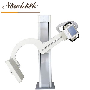
When the anatomy is longer than the detector, a room has three options. Understanding all three sharpens the buying question:
| Route | How it works | What it demands from the room |
|---|---|---|
| Single large-field exposure | A large detector (for example, a 17″ × 17″ panel) captures the region of interest in one shot | Detector size is the hard limit — a full femur or full spine still exceeds one field; suitable for joints and segments, not full-length studies |
| Segmented exposures + software stitching | The system acquires two or more overlapping exposures along the anatomy, then the software merges them into one full-length image | Constant source-to-image distance between segments, a precise overlap, and either a smoothly traveling arm or a moving detector carriage; the software must be documented, not implied |
| Legacy long-film / elongated receptor | Computed or film-era technique with a long cassette | Largely displaced by DR; relevant only if you are inheriting an older room |
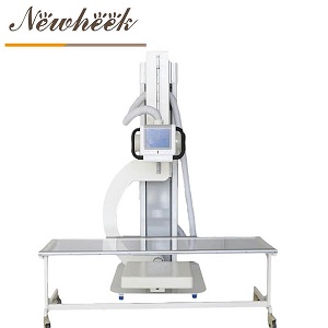
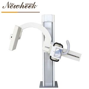
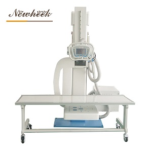
The middle route is the one 2026 procurement conversations are about. And it is where the U-arm earns its keep: the tube and detector hang on one rigid arm, so the source-to-image geometry that stitching software assumes — same distance, same beam-detector axis, no skew between segments — is preserved by construction, not by the technologist’s vigilance.
Published U-arm platform figures (the same motion map our sickle-arm explainer walks through) translate directly into stitching capability:
None of this means every U-arm stitches. The travel must be smooth and adequately slow at acquisition, the overlap must be controlled, and the stitching software itself is a separate, licensable capability. Treat the structure as the enabler, and the software as the product feature you verify in writing.
Stitched long-bone work and weight-bearing work share a room. The U-arm’s rotation range (published at −30° to +120° around the column) puts the detector against the wall-side of the room for upright spine and lower-limb studies, then swings the same tube-detector pair over the table for supine work — the patient stays put between projections. For patients who cannot stand at all, the same room logic is covered in our guide to wheelchair, seated and mobility-limited workflows.
Two practical notes for standing orthopedic work:
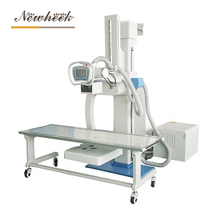
Stitching capability is bought, documented and tested — or it disappoints. Copy these into your RFQ:
For the room-side prerequisites that stitched upright work depends on — ceiling height, floor loading, power — our U-arm room requirements checklist covers the layout before you order. Configuration options are on the multifunctional U-arm radiography system page.
The single-arm structure provides the geometric preconditions — constant SID, fixed tube-detector alignment, straight-line travel. Whether a specific configuration offers stitching software, and for which anatomy, depends on the supplier’s offering. Ask for the capability in writing and include a stitched study in the acceptance test.
Both exist. Some systems stitch from manually or automatically positioned overlapping exposures with software on the acquisition workstation; others add motorized detector or arm movement for the acquisition pass. Confirm which one a quotation refers to, because reproducibility and price differ.
Alignment under load is the clinical question: joint space, limb alignment and spinal posture change between lying and standing. That is why suppliers of orthopedic DR workflows make standing positioning an explicit feature — the image that answers the surgical question is usually taken on the patient’s feet.
A stitched study uses multiple exposures, so the total is higher than one plain film of the same region — but each segment can be collimated tightly to its own field, and a first-pass diagnostic stitched study avoids repeating the whole series. Collimation discipline and a low repeat rate are the practical dose controls.
That is the platform’s core promise: one rigid tube-detector pair sweeping from an upright wall-side position down over the table. Which exam protocols — including any stitching applications — are validated for your configuration should be confirmed with the manufacturer before purchase.
Planning an orthopedic-leaning room — standing views, long-bone segments, stitching on the shortlist? Tell us your examination mix and we will map it onto a configuration and a written capability list. Reach us at admin@newheek.cn or WhatsApp +86 19062611512, or send the inquiry form in our Contact Us module and our product manager will respond within 24 hours on working days.
Related reading: U-arm DR for lumbar spine imaging · What does a U-arm X-ray system cost in 2026?
Tel: +86 19062611512
Email : admin@newheek.cn
Company : Weifang Newheek Electronic Technology Co., Ltd.