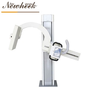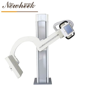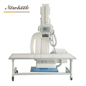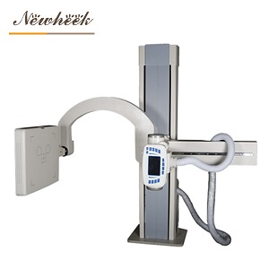

The sickle-arm DR filming machine has developed by leaps and bounds in recent years. The pictures taken are delicate, high-definition, high-resolution, and good image effects. It is more convenient for doctors to judge the patient’s condition, and it uses a lower dose of radiation to obtain high-quality images. The advantages of reduced skin dose and minimal X-ray damage to doctors and patients have made the sickle arm DR film machine affirmatively recognized in the radiology industry.
Why is there such a big change in the function of the sickle arm DR filming opportunity? Of course, these factors are determined by the different hardware and software used. Next, I will take you to take a look at what equipment the sickle arm DR filming machine produced by our Newheek is composed of!
1. Toshiba flat panel detector: The size of the collected image can reach 43×43cm (17×17 inches).
2. Acquisition software: HR high-resolution mode: suitable for observation of limbs and details; HE high-efficiency mode: suitable for thick tissues such as lumbar spine and chest. Friendly Chinese user interface, easy to operate, easy to achieve image acquisition, organization balance, contrast enhancement, edge enhancement, image smoothing/sharpening, filtering and other image processing functions, standard DICOM interface, strong network compatibility.
3. Dell acquisition workstation: dual-core mode, 2G memory, 320G hard drive, 19-inch display.
4. High voltage generator.
5. Toshiba tube.
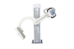
6. Intelligent UC arm rotation angle: -30 degrees to 120 degrees
7. Four-wheel mobile photography bed
8. Manual shutter
9.17X17 inch grid
10. High voltage cable with 75KV withstand voltage
11. Post-processing workstation: dual-core mode, 2G memory, 320G hard disk, printing graphic reports, medical record templates and other functions, HP laser printer
12. Professional console: equipped with power supply, network port and other interfaces
Tel: +86 17616362240
Email : newheek1999@outlook.com
Company : Weifang Newheek Electronic Technology Co., Ltd.